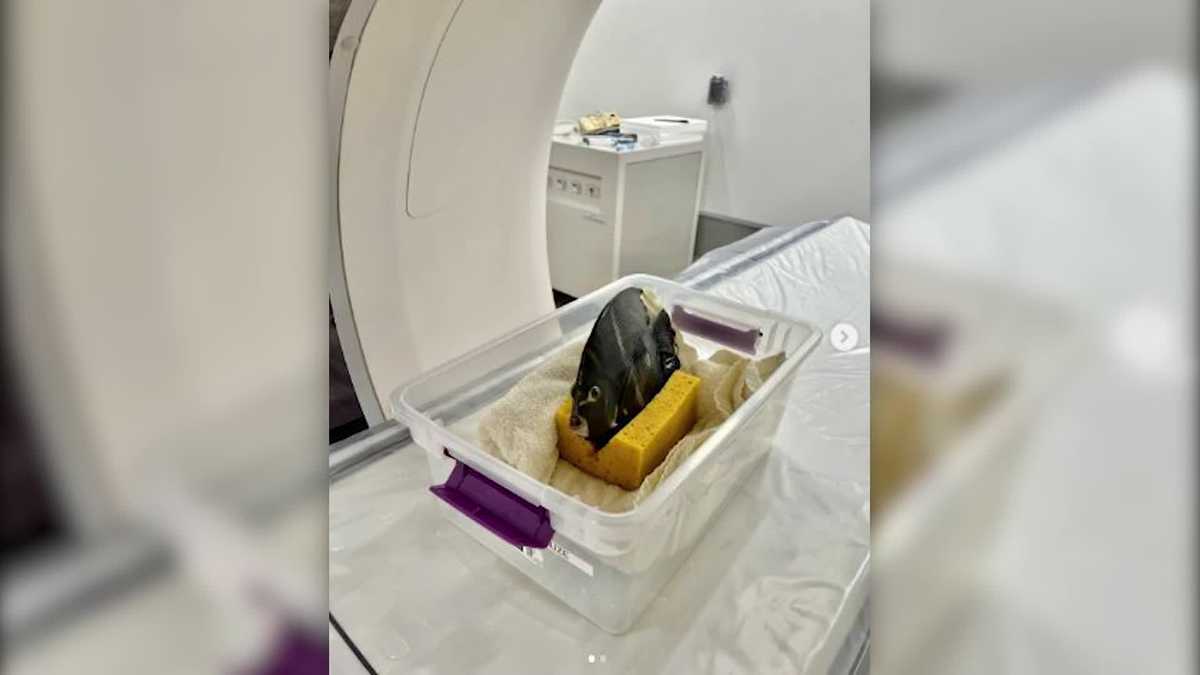
Denver Zoo Fish CT Scan
A CT scan, or computed tomography scan, is a medical imaging technique that uses X-rays and computer processing to create detailed cross-sectional images of internal body structures. It is a non-invasive procedure that is often used to diagnose and monitor a wide range of medical conditions, including cancer, heart disease, and lung disease. CT scans can also be used to guide biopsies and other medical procedures.At the Denver Zoo, CT scans are used to diagnose and treat a variety of health problems in animals. For example, CT scans can be used to:
- Detect and diagnose cancer
- Evaluate the extent of injuries
- Plan for surgery
- Monitor the response to treatment
CT scans are a safe and effective way to diagnose and treat a variety of health problems in animals. They are a valuable tool for veterinarians, and they can help to improve the quality of life for animals.
Denver Zoo Fish CT Scan
The Denver Zoo uses CT scans to diagnose and treat a variety of health problems in animals. CT scans are a valuable tool for veterinarians, as they provide detailed images of the internal anatomy of animals. This information can help veterinarians to make accurate diagnoses and develop effective treatment plans.
- Non-invasive: CT scans are a non-invasive procedure, meaning that they do not require surgery or anesthesia.
- Detailed images: CT scans provide detailed images of the internal anatomy of animals, which can help veterinarians to make accurate diagnoses.
- Variety of uses: CT scans can be used to diagnose and treat a variety of health problems in animals, including cancer, heart disease, and lung disease.
- Safe and effective: CT scans are a safe and effective way to diagnose and treat health problems in animals.
- Planning for surgery: CT scans can be used to plan for surgery, by providing detailed images of the surgical area.
- Monitoring treatment: CT scans can be used to monitor the response to treatment, by providing images of the treated area over time.
- Emergency care: CT scans can be used in emergency care situations, to quickly and accurately diagnose life-threatening injuries.
- Research: CT scans can be used for research purposes, to study the anatomy of animals and to develop new treatments for diseases.
- Education: CT scans can be used for educational purposes, to teach students about the anatomy of animals and the use of CT scans in veterinary medicine.
CT scans are a valuable tool for veterinarians, and they can help to improve the quality of life for animals. They are a safe and effective way to diagnose and treat a variety of health problems in animals, and they can be used for a variety of purposes, including emergency care, research, and education.
Non-invasive
This is an important consideration for zoo animals, as anesthesia can be risky and stressful. CT scans allow veterinarians to get detailed images of the internal anatomy of animals without having to put them under anesthesia. This is especially important for animals that are already sick or injured, as anesthesia can further compromise their health.
For example, in 2019, the Denver Zoo used a CT scan to diagnose a cancerous tumor in a 22-year-old orangutan named Budi. The CT scan allowed the zoo's veterinarians to determine the size and location of the tumor, and to develop a treatment plan. Budi underwent surgery to remove the tumor, and he is now doing well.
CT scans are a valuable tool for veterinarians, as they provide a safe and effective way to diagnose and treat a variety of health problems in animals. The non-invasive nature of CT scans makes them especially useful for zoo animals, as it allows veterinarians to get detailed images of their internal anatomy without having to put them under anesthesia.
Detailed images
CT scans provide detailed images of the internal anatomy of animals, which can help veterinarians to make accurate diagnoses. This is especially important for zoo animals, as many of them are exotic species that veterinarians may not be familiar with. CT scans can help veterinarians to identify diseases and injuries that would be difficult or impossible to diagnose with other methods.
- Early detection: CT scans can help veterinarians to detect diseases and injuries at an early stage, when they are more likely to be treatable. For example, CT scans can be used to detect cancer tumors, heart disease, and lung disease in animals.
- Accurate diagnosis: CT scans can help veterinarians to make more accurate diagnoses by providing detailed images of the internal anatomy of animals. This can help to rule out other possible diseases and injuries.
- Treatment planning: CT scans can help veterinarians to plan for surgery and other treatments by providing detailed images of the surgical area. This can help to ensure that the surgery is performed safely and effectively.
- Monitoring treatment: CT scans can be used to monitor the response to treatment by providing images of the treated area over time. This can help veterinarians to determine if the treatment is working and to make adjustments as needed.
CT scans are a valuable tool for veterinarians, as they provide a safe and effective way to diagnose and treat a variety of health problems in animals. The detailed images that CT scans provide can help veterinarians to make accurate diagnoses and to develop effective treatment plans.
Variety of uses
CT scans are a valuable tool for veterinarians, as they can be used to diagnose and treat a wide variety of health problems in animals. This is especially important for zoo animals, as many of them are exotic species that veterinarians may not be familiar with. CT scans can help veterinarians to identify diseases and injuries that would be difficult or impossible to diagnose with other methods.
For example, at the Denver Zoo, CT scans have been used to diagnose and treat a variety of health problems in animals, including:
- Cancer: CT scans can be used to detect and diagnose cancer tumors in animals. This information can help veterinarians to determine the stage of the cancer and to develop a treatment plan.
- Heart disease: CT scans can be used to evaluate the heart and blood vessels of animals. This information can help veterinarians to diagnose heart disease and to monitor the response to treatment.
- Lung disease: CT scans can be used to evaluate the lungs of animals. This information can help veterinarians to diagnose lung disease and to monitor the response to treatment.
CT scans are a safe and effective way to diagnose and treat a variety of health problems in animals. The variety of uses for CT scans makes them a valuable tool for veterinarians, and they can help to improve the quality of life for animals.
Safe and effective
CT scans are a safe and effective way to diagnose and treat health problems in animals. This is especially important for zoo animals, as many of them are exotic species that veterinarians may not be familiar with. CT scans can help veterinarians to identify diseases and injuries that would be difficult or impossible to diagnose with other methods.
For example, at the Denver Zoo, CT scans have been used to diagnose and treat a variety of health problems in animals, including cancer, heart disease, and lung disease. In one case, a CT scan was used to diagnose a cancerous tumor in a 22-year-old orangutan named Budi. The CT scan allowed the zoo's veterinarians to determine the size and location of the tumor, and to develop a treatment plan. Budi underwent surgery to remove the tumor, and he is now doing well.
CT scans are a valuable tool for veterinarians, as they provide a safe and effective way to diagnose and treat a variety of health problems in animals. The safe and effective nature of CT scans makes them especially useful for zoo animals, as it allows veterinarians to get detailed images of their internal anatomy without having to put them under anesthesia.
Planning for surgery
Planning for surgery is a critical step in ensuring a successful outcome. CT scans play a vital role in surgical planning by providing detailed images of the surgical area. This information allows surgeons to visualize the anatomy of the area to be operated on, identify potential risks, and plan the best approach for the surgery.
For example, at the Denver Zoo, CT scans are routinely used to plan for surgery in a variety of animals. In one case, a CT scan was used to plan for surgery to remove a cancerous tumor from a lion. The CT scan allowed the surgeons to determine the size and location of the tumor, and to plan the best approach for removing it. The surgery was successful, and the lion is now cancer-free.
CT scans are a valuable tool for surgeons, as they provide detailed images of the surgical area. This information allows surgeons to plan for surgery more effectively, which can lead to better outcomes for patients.
Monitoring treatment
Monitoring the response to treatment is essential to ensure that the treatment is effective and that the patient is recovering as expected. CT scans play a vital role in monitoring treatment by providing detailed images of the treated area over time. This information allows doctors to assess the progress of the treatment and to make any necessary adjustments.
- Assessing progress: CT scans can be used to assess the progress of treatment by comparing images taken before and after treatment. This information can help doctors to determine if the treatment is working and if the patient is responding as expected.
- Identifying complications: CT scans can also be used to identify any complications that may arise during treatment. For example, CT scans can be used to detect infections, bleeding, or other problems that may require additional treatment.
- Adjusting treatment: The information provided by CT scans can be used to adjust treatment as needed. For example, if a CT scan shows that the treatment is not working as expected, the doctor may adjust the dosage or frequency of the treatment.
CT scans are a valuable tool for monitoring the response to treatment in animals at the Denver Zoo. For example, CT scans have been used to monitor the response to treatment in animals with cancer, heart disease, and lung disease. In one case, a CT scan was used to monitor the response to treatment in a lion with a cancerous tumor. The CT scan showed that the tumor was shrinking in response to treatment, and the lion is now cancer-free.
CT scans are a safe and effective way to monitor the response to treatment in animals. They provide detailed images of the treated area over time, which allows doctors to assess the progress of the treatment and to make any necessary adjustments. This information can help to ensure that animals receive the best possible care and that they recover as quickly as possible.
Emergency care
CT scans play a vital role in emergency care, as they can quickly and accurately diagnose life-threatening injuries. This is especially important for zoo animals, as they may not be able to communicate their symptoms to their caregivers. CT scans can help veterinarians to identify and treat injuries such as bleeding, fractures, and organ damage.
- Rapid diagnosis: CT scans can be performed quickly, which is essential in emergency situations. This allows veterinarians to quickly diagnose life-threatening injuries and to start treatment immediately.
- Accurate diagnosis: CT scans provide detailed images of the internal anatomy of animals, which allows veterinarians to accurately diagnose injuries. This information can help veterinarians to determine the best course of treatment.
- Treatment planning: CT scans can be used to plan for surgery and other treatments by providing detailed images of the injured area. This information can help veterinarians to ensure that the surgery is performed safely and effectively.
CT scans are a valuable tool for veterinarians in emergency care situations. They can help veterinarians to quickly and accurately diagnose life-threatening injuries and to start treatment immediately. This can help to improve the chances of survival for zoo animals.
Research
CT scans are a valuable tool for researchers, as they provide detailed images of the internal anatomy of animals. This information can be used to study the anatomy of animals, to develop new treatments for diseases, and to improve the care of animals in zoos.
For example, at the Denver Zoo, CT scans have been used to study the anatomy of a variety of animals, including fish. This information has been used to develop new treatments for diseases that affect fish, such as swim bladder disease and ich. CT scans have also been used to improve the care of animals in the zoo, by providing veterinarians with detailed images of the internal anatomy of animals. This information can be used to diagnose diseases, plan for surgery, and monitor the response to treatment.
The research that is conducted using CT scans at the Denver Zoo is helping to improve the care of animals in zoos and to develop new treatments for diseases that affect animals. This research is also helping to advance our understanding of the anatomy of animals, which is important for the conservation of endangered species.
Education
CT scans are a valuable tool for education, as they provide detailed images of the internal anatomy of animals. This information can be used to teach students about the anatomy of animals and the use of CT scans in veterinary medicine.
At the Denver Zoo, CT scans are used to teach students about the anatomy of a variety of animals, including fish. This information is used to develop educational programs for students of all ages. For example, the zoo's "Animal Anatomy Adventure" program uses CT scans to teach students about the anatomy of different animals, including fish. This program has been used to teach thousands of students about the anatomy of animals and the use of CT scans in veterinary medicine.
Education is an important component of the Denver Zoo's mission. The zoo is committed to providing educational opportunities for students of all ages. CT scans are a valuable tool for education, as they provide detailed images of the internal anatomy of animals. This information can be used to teach students about the anatomy of animals and the use of CT scans in veterinary medicine.
FAQs about Denver Zoo Fish CT Scan
This FAQ section provides answers to common questions and concerns about the use of CT scans on fish at the Denver Zoo.
Question 1: What is a CT scan?
A CT scan, or computed tomography scan, is a medical imaging technique that uses X-rays and computer processing to create detailed cross-sectional images of internal body structures. CT scans are a non-invasive procedure that is often used to diagnose and monitor a wide range of medical conditions, including cancer, heart disease, and lung disease.
Question 2: Why are CT scans used on fish at the Denver Zoo?
CT scans are used on fish at the Denver Zoo to diagnose and treat a variety of health problems, including cancer, heart disease, and lung disease. CT scans can also be used to plan for surgery, monitor the response to treatment, and conduct research.
Question 3: Are CT scans safe for fish?
Yes, CT scans are safe for fish. The Denver Zoo uses a low-dose CT scanner that is specifically designed for use on animals. The radiation exposure from a CT scan is minimal, and it is far below the level that would be harmful to fish.
Question 4: How are fish prepared for a CT scan?
Fish are prepared for a CT scan by being anesthetized. This ensures that the fish remain still during the scan. Once the fish is anesthetized, it is placed on the CT scanner table and the scan is performed.
Question 5: How long does a CT scan take?
The length of a CT scan varies depending on the size of the fish and the area being scanned. However, most CT scans take between 5 and 15 minutes.
Question 6: What are the benefits of using CT scans on fish?
CT scans provide a number of benefits for fish, including:
- Early detection of disease
- Accurate diagnosis
- Planning for surgery
- Monitoring the response to treatment
- Research
CT scans are a valuable tool for veterinarians at the Denver Zoo. They provide detailed images of the internal anatomy of fish, which can help veterinarians to diagnose and treat a variety of health problems. CT scans are safe, effective, and provide a number of benefits for fish.
For more information about CT scans at the Denver Zoo, please visit our website.
Denver Zoo Fish CT Scan Tips
CT scans are a valuable tool for veterinarians at the Denver Zoo. They provide detailed images of the internal anatomy of fish, which can help veterinarians to diagnose and treat a variety of health problems. Here are a few tips to help you get the most out of your fish's CT scan:
Tip 1: Prepare your fish for the scan. Fish should be fasted for 24 hours prior to the scan. This will help to ensure that the fish's stomach and intestines are empty, which will provide better images.
Tip 2: Bring your fish's medical history to the scan. This will help the veterinarian to interpret the results of the scan and to make a diagnosis.
Tip 3: Be present for the scan. This will allow you to ask the veterinarian any questions that you may have and to learn more about the results of the scan.
Tip 4: Follow the veterinarian's instructions after the scan. The veterinarian may recommend that you follow up with your fish's care at home. Be sure to follow the veterinarian's instructions carefully to ensure that your fish makes a full recovery.
Tip 5: Be patient. It may take some time for the veterinarian to interpret the results of the scan and to make a diagnosis. Be patient and allow the veterinarian to take their time.
CT scans are a safe and effective way to diagnose and treat a variety of health problems in fish. By following these tips, you can help to ensure that your fish's CT scan is a success.
For more information about CT scans at the Denver Zoo, please visit our website.
Conclusion
CT scans are a valuable tool for veterinarians at the Denver Zoo. They provide detailed images of the internal anatomy of fish, which can help veterinarians to diagnose and treat a variety of health problems. CT scans are safe, effective, and provide a number of benefits for fish.
The Denver Zoo is committed to providing the best possible care for its animals. CT scans are an important part of that commitment. By using CT scans, the Denver Zoo can provide its animals with the best possible diagnosis and treatment.
:max_bytes(150000):strip_icc():focal(421x139:423x141)/fish-getting-ct-scan-denver-zoo-tout-083123-a213cebb76014c148b3499f17f7a2161.jpg)
Detail Author:
- Name : Dr. Brennon Boyer Sr.
- Username : meredith.goodwin
- Email : raquel.weimann@schinner.com
- Birthdate : 1990-11-05
- Address : 29514 Ruthe Parks Suite 275 East Stanton, DE 63674
- Phone : +1-501-603-3497
- Company : Kessler, Murray and Carroll
- Job : Service Station Attendant
- Bio : Ipsa optio sequi corporis quo error animi sint. Ut sit at distinctio facere similique. Sint sunt doloremque pariatur.
Socials
linkedin:
- url : https://linkedin.com/in/willis.auer
- username : willis.auer
- bio : Ea rerum hic laudantium itaque.
- followers : 5556
- following : 2845
instagram:
- url : https://instagram.com/auer1987
- username : auer1987
- bio : Nulla quo modi asperiores nam eos. Non consectetur minima omnis.
- followers : 5651
- following : 2497
twitter:
- url : https://twitter.com/willis8795
- username : willis8795
- bio : Asperiores beatae possimus adipisci velit. Odit perspiciatis sequi quod quis quaerat. Molestiae dolore veritatis qui quo possimus.
- followers : 1716
- following : 1837
tiktok:
- url : https://tiktok.com/@auerw
- username : auerw
- bio : Fuga eaque nihil cumque dolores quia voluptatem.
- followers : 3511
- following : 2928